
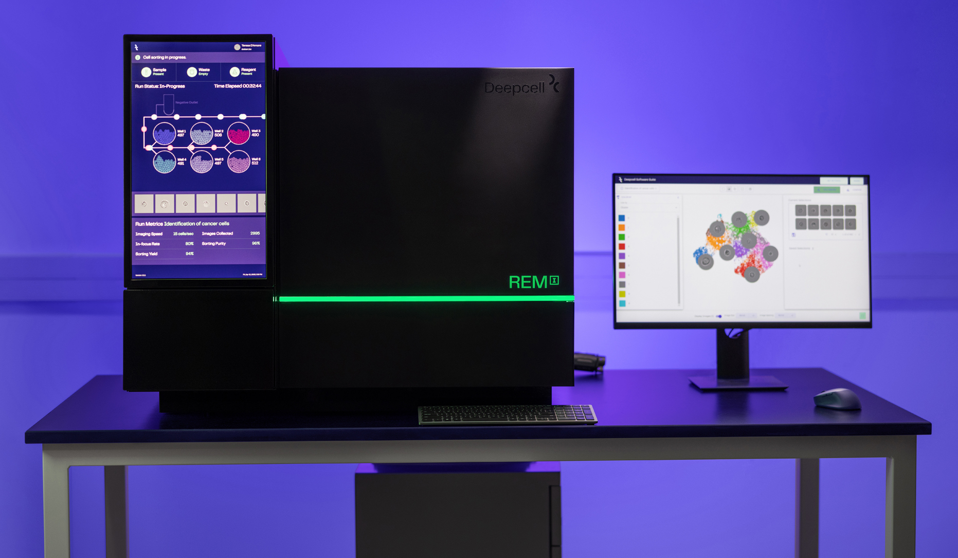
Experience the REM-I Platform
platform
Introducing the REM-I Platform
Methodologies to assess and characterize cell morphology, which can be highly indicative of phenotype and function, have been limited to either imaging or sorting with labels, until now.
The REM-I Platform takes the best of these worlds to provide imaging of single cells and label-free sorting in one platform leveraging AI.

Real-time characterization with
the Human Foundation Model
REM-I imaging and sorting consumables
REM-I instrument
Axon data suite
True imaging
High-resolution imaging of single cells
The REM-I instrument takes high-speed, high-resolution brightfield images of single cells to capture information about morphology.
Gentle sorting
6-way, label-free sorting
A label-free workflow and gentle microfluidics means the cells you sort are minimally perturbed and viable for downstream analysis.
High-dimensional output
Powered by Deepcell’s Human Foundation Model
The Human Foundation Model assesses many dimensions of cell morphology for a high-dimensional characterization of each cell.
Powerful data suite
Real-time analysis of single cell morphology
Store, visualize, and analyze single cell image and high-dimensional cell morphology data with the Axon data suite.
A single, streamlined workflow

Prepare cells into single cell suspension

Image single cells in flow

Characterize cells in real-time with the HFM

Analyze data and optionally collect cell populations of interest for further analysis
Applications
Explore drivers of disease state, drug effects, developmental processes, perturbations, and more using high-dimensional morphology as its own modality.

Cancer research
Detect disease and identify morphological heterogeneity within and between cancer samples
Developmental biology
Classify cells by type and activation state with morphology as a marker
Cell & gene therapy
Predict the effect of genetic perturbations on phenotype with morphology
Drug & functional screening
Screen for cell health and morphological impacts of drug treatments
Dr. Peter van der Spek
Professor of Clinical Bioinformatics, Pathology“The biggest advantage is that we don’t have to do staining – we simply push our cells/samples through REM-I and by the end of the day you have the high-dimensional morphology data of 100,000 or more cells.”
Dr. Peter van der Spek - Professor of Clinical Bioinformatics, Pathology
Dr. Andrew Filby
Professor, Flow Cytometry Core Director, Newcastle University“There are different methods for characterizing cells at the single cell level, for example flow cytometry or imaging cells in flow, but they perturb the cells and don’t have the ability to sort for downstream analysis like REM-I does.”
Dr. Andrew Filby - Professor, Flow Cytometry Core Director, Newcastle University
Dr. Andy Tsai
Postdoctoral Scholar, Neurology, Stanford University“We’ve seen first hand how the REM-I platform can validate and speed up the discovery and diagnosis of disease.”
Dr. Andy Tsai - Postdoctoral Scholar, Neurology, Stanford University
Dr. Jennifer Yokoyama
Associate Professor, Neurology, USCF Weill Institute for Neurosciences“Mopholomics represents the natural next step to learn more about diseases, their biology, and identify novel targets for disease treatment in an unbiased fashion.”
Dr. Jennifer Yokoyama - Associate Professor, Neurology, USCF Weill Institute for Neurosciences

Request pricing